1. The
Full Mouth Reconstruction using Magnetic Attachments
Nakamura Y., Shoji K., Ando A. , Tanaka T.1 , Okada M.1 ,
Imaoka S., Ohno Y. and Tanaka Y.
Removable Prosthodontics, School of Dentistry, Aichi-Gakuin University
1Department of Dental Laboratory , Aichi - Gakuin University Dental
Hospital
Introduction
A magnetic attachment is a
device using magnetic attractive force to provide and assist in the retention
of dentures. Dentures using these special attachments have been well received
by patients and treating dentists. The purpose of this paper is
the case presentation of a patient who presented with chief complaints of
aesthetic dissatisfaction and inability to chew. This patient underwent full
mouth reconstruction using magnetic attachments as retaining elements. The
following is a summary of treatment completed.
Clinical History
The patient was a 42-year-old female with
chief complaints of aesthetic dissatisfaction and masticatory dysfunction. The
patient had received implants on the right lower molar region in 1995, but they
were failed after 3 years. The edentulous upper and lower molar regions were
left untreated for many years, resulting in a distorted plane of the occlusion
and a decrease in occlusal vertical dimension. Although the patient visited
several general practitioners and municipal hospitals to seek aesthetic
correction, she was dissatisfied with the treatment results, and then consulted
with our department.
Initial Status
The patient had Kennedy Class II relationship
in the upper and lower arches. There was severe molar occlusal destruction with
a occlusal plane discrepancy. Also present were areas of root fracture, failing
restorations with ill-fitting margins, and poor periodontal tissue health.
There was severe redness and swelling of periodontal tissues around the right
upper lateral incisor, and right lower first premolar areas. Radiographic
examination demonstrated endodontic problems of apical radiolucencies and poor
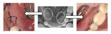
endodontic fills in other areas. Clinical examination revealed the bone torus
which can be an obstacle to the restorative treatment (Fig. 1).
Treatment Procedure
1 .
Exploration
Fig
. 2 Face-bow transfar and Wax up for treatment
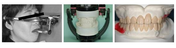
The mounted study casts
were made and mounted on an articulator using face-bow transfer procedure. The
diagnostic wax-up was performed to show an anticapated result and establish a
restorative treatment plan showing space and dental relationship problems that
might exist. The diagnostic modeling provides excellent material for patient
education and demonstration (Fig. 2).
Initial
caries control and treatment of endodontic etiology., Extractions if necessary.
2 .
Temporary Restoration and Initial Treatment Denture
The quality of the existing
restorations caused a poor occlusal and periodontal environment. These were
removed. Temporary restorations were placed to secure the temporary masticatory
function, pronunciation, and to evaluate ane initial esthetic result, and to
provide for initial periodontal and occlusal treatments stability. Temporary
restorations were fabricated based on the diagnostic wax-up. In the edentulous area, treatment
denture was placed to improve the occlusal support (Fig. 3).
![]()

3 .
Provisional Restoration
After the completion of the
initial periodontal treatment, resin cores with fiber posts were placed in the
remaining teeth to avoid dental fracture. A provisional restoration with
improved esthetics and occlusal height, and adjusted occlusal vertical
dimension from the temporary restoration was fabricated (Fig. 4).
4. Surgical Treatment
Maxillary bony exostosis
that may interfere with final denture fabrication was surgically resected (Fig.
5).
![]()
5. Preparation
Fig
. 6 Preparation for Abutment Tooth
To
protect vital abutment teeth, preparation was performed in 3 steps with the
time interval to promote the formation of the secondary dentin that protects
the pulp.(Fig. 6)
6. Final Provisional Restoration
A final provisional
restoration with improved occlusion, periodontal environment and esthetics was
fabricated and presented for approval by the patient. .
7.
Design of Final Restoration
Table
. 1 Upper Design
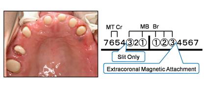
Maxillary denture design
includes an extracoronal magnetic attachment on the distal surface of the left
upper canine, slit on the distal surface of the right upper canine, and metal
crowns on the right upper second premolar and first molar (Table 1).
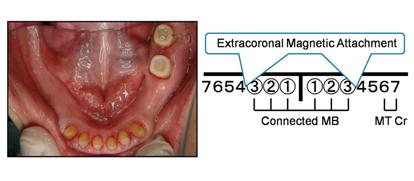
Mandibular denture
design includes an extracoronal magnetic attachment on the distal surface of
the canines, and metal crowns on the left first and second molars (Table 2).
![]()
8.
Maxillo-mandibular Relationships and Articulator Mounting.
Cross mount transfer technique
was used to preserve the original maxillo-mandibular cast relations and
mounting. This procedure also preserves original diagnostic occlusal design and
vertical dimension of the provisional restoration to the final restoration
(Fig. 7). Since provisional and work models are transferred on the same axis,
these models can also be compared during fabrication of the 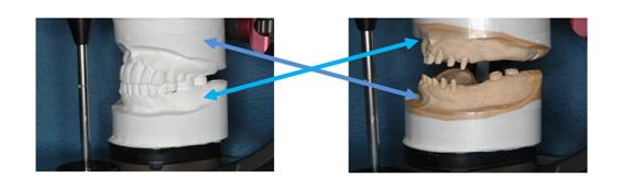 final restoration to use as a
reference to the opposing arch and teeth. This method simplifies the
final restoration to use as a
reference to the opposing arch and teeth. This method simplifies the
![]()
fabrication procedures
such as wax-up.
9.
Trial Fitting Procedures
Trial fitting of the metal
work and metal crowns was performed. Transfer impression was taken following
the conventional methods after try fitting, followed by the fabrication and try
fitting of the wax denture.
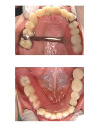
10.
Final Restoration
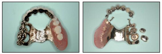
Fig. 8 shows upper and Fig.
9 shows lower final restorations.
Fig
. 8 Upper Final Restoration
Fig
. 9 Lower Final Restoration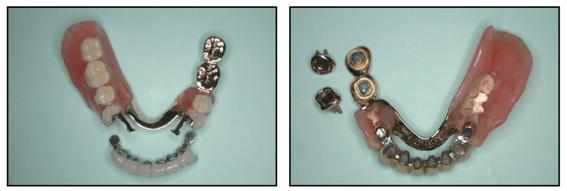
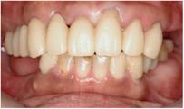
Discussion
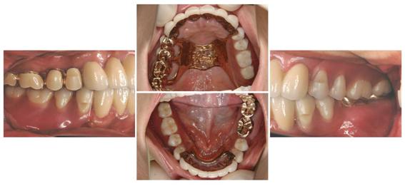
The patient was
satisfied with the esthetic results of the final restoration with magnetic
attachments. The retention obtained
with the attachments achieved highly satisfactory functional results. The
patient was satisfied with the prosthetic retentive force (Fig. 10).
![]()
The postoperative course
has been uneventful. However, the
design of a final restoration is complex as the ideal combination of esthetics
and functionality is difficult to obtain. It is important that regular
maintenance is obtained. (Fig. 11).
References
1. Gillings, B. R. D.: Magnetic retention
for complete and partial overdentures, Part. J. Prosthet. Dent., 45(5):
484-491, 1981.
2. Jackson,T.R.: The application of rare
earth magnetic retention toosseointegrated implants. Int. J. Oral & Maxill.
Imp., 1:81-92, 1986.
3. Tanaka, Y.: Dental Magnetic Attachment,
Q & A, Ishiyaku Publishers, Inc. (Tokyo),
1995.
4. Mizutani, H., Ishihata, N. and Nakamura,
K.: Removable partial denture used the magnetic attachment, Quintessence
Publishing Co., Ltd. (Tokyo), 1994.