14. Influence of the magnetic attachment on blood flow to the surrounding oral mucosa
Fukuzawa R., Hasegawa N., Syouji K., Ando A., Nakamura Y. and Tanaka Y.
The First Department of Prosthodontics, School of Dentistry , Aichi-Gakuin University
Introduction
The use of a magnet and magnetism has been drawing attention in the modern society, and they have been widely applied in the medical field in recent years. It is a major research task for us to confirm the safety of a magnet in the human body. Several studies including cytotoxicity test and corrosion test have been conducted. Several reports have been suggested that the magnetism increases blood flow, but not solid data exist regarding a magnetic attachment. The present paper reports interesting findings that we obtained from the detailed and clear observation of the influence of magnetic field on human blood flow.
Objective
The following three experiments were conducted to confirm the influence of magnetic field on human blood flow of the surrounding oral mucosa.
Materials and Methods
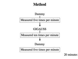
1. Experiment 1
1)Sample: GIGAUSS D 1000 (GC)
2)Leakage of magnetic fields: 150 mT, ∅4.9 x 1.3 mm
3)Measuring device: Laser blood flowmeter ALF 21D (advance)
4)Subjects: Eight healthy dentate subjects ♂ (20-30 years of age)
5)Measurement points and
placement of samples(Fig. 1)
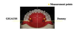
Fig. 1 Measurement
points and placement of samples
6)The position: Decline position
7)Duratin of measurement: Five minutes at both points
8)Control: Value at five
minutes before each sample fixation
2. Experiment 2
1)Sample: GIGAUSS D 1000 (GC)
2)Measuring device: Laser Doppler perfusion imaging periscan PIM II (Perimed)
3)Subjects: Four healthy dentate subjects ♂ (20-30 years of age)
4)Measurement points and
placement of samples( Fig. 2)
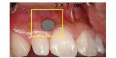
Fig. 2 Measurement
points and placement of samples
5)Measurement procedure
3. Experiment 3
1)Sample: Nd-Fe magnet
2)Leakage of magnetic fields: 300 mT, ∅ 20.0 x 5.0 mm
3)Measurement points and
placement of samples (Fig. 3)
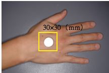
Fig. 3 Measurement points and placement of samples
4)Measuring device, Subjects: The measurement was carried out following the same procedure as in Experiment 2.
Results
1. Experiment 1
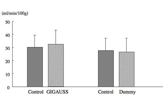
Fig. 4 Mean values and standard deviation of blood flow in 8 subjects
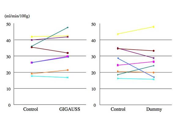 Fig. 5 Blood flow of each subject
2. Experiment 2
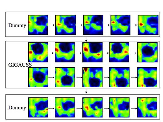
Fig. 6 Typical images of blood flow in the alveolar mucosa
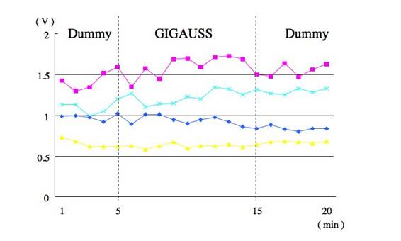
Fig. 7 Change with time in the mean value within the measurement range of each subject
3. Experiment 3
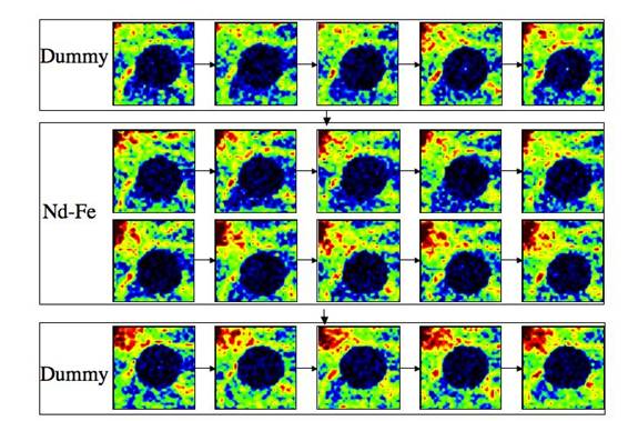
Fig. 8 Typical images of blood flow in the dorsal surface of the hand

Fig. 9 Change with time in the mean value within the measurement range of each subject
Discussions
In the Experiment 1, we continuously measured at one specific point to observe the influence of the magnetic field. The results showed little influence of magnetic attachments on blood flow of the surrounding tissue. However, we speculated that different blood flow with each individual in the different regions, and susceptibility to the course of anatomical blood vessels may have resulted in the data variation, and conducted the Experiment 2 using a laser Doppler perfusion imaging periscan PIM II. This device enables intermittent but extensive measurement. Since the influence of the magnetic field was not observed in this experiment, we conducted Experiment 3 using Ne-Fe magnet with a large magnetic field as a sample, but no significant difference was observed between the results obtained from the Experiment 2 and 3. These results suggested that a magnetic attachment is not invasive to the human body in terms of blood circulation.
Conclusions
No change was observed in blood flow of the surrounding tissue of a magnetic attachment.
References
1. Tanaka, Y.: Dental Magnetic Attachment - new prothodontic treatment used the magnet -, Ishiyaku Publishers, Inc. (Tokyo), 1992.
2. Tanaka, Y.: Dental Magnetic Attachment, Q&A, Ishiyaku Publishers, Inc. (Tokyo), 1995.
3. Nakamura, Y.: Stress analysis of overlay denture and a magnetic attachment using finite element method. J Jpn Prosthodont Soc, 42: 234-245, 1998.