6. The effect of tooth connection with a magnetic extracoronal attachment using the Three Dimensional Finite Element Method
Ando A. , Kumano H. , Miyata T. , Masuda T. , Nakamura Y. and Tanaka Y.
The First Department of Prosthodontics , School of Dentistry , Aichi – Gakuin University
Introduction
The magnetic attachment design has been well received by patients and dentists for both functional advantage and the improvement of aesthetic result. A conventional magnetic attachment is generally designed for use with non-vital teeth due to space considerations, but we have developed an extracoronal magnetic attachment which can be applied with both vital and non-vital teeth. It is a retainer design that is considered stress releasing upon clinical application due to the lateral side play and give between the magnetic keeper and magnet. A magnetic attachment provides support and retention elements, but has no bracing element for horizontal force control. Therefore, in applications as a extracoronal attachment, the bracing requirement in the design is overcome by using an engaging arm, and interlock or addition of a slit into the attachment. In short, the denture and abutment tooth work together in unison to serve as a more rigid retainer.
Unilateral treatment of a distal extension partial denture is considered to be contraindicated since the functional pressure applied to the denture is directly transmitted to the most distal abutment tooth due to the difference in the amount of tissue displacement between the periodontal membrane and the mucosa. However, there are patient requests for unilateral prosthetic partial designs and clinicians may be obliged to consider these alternative designs for restorative prosthetic treatment using these designs. In these treatment conditions, a lack of attention to proper design and the condition of abutment teeth could lead to the root fracture or attachment breakage.
Objective
In the clinical treatment situations, the preservation of remaining periodontal tissue support is made by increasing the number of prosthetic abutment tooth supports. However, the decision in the number of interconnected abutment teeth is made based on the dentists’ clinical experience, and evaluations of treatment conditions. There may be no solid evidence to base a certain treatment decision regarding prosthetic design. In this present study, we investigated the effect of abutment teeth connection with a magnetic extracoronal attachment using the finite element method.
Materials and Methods
1. Analysis model
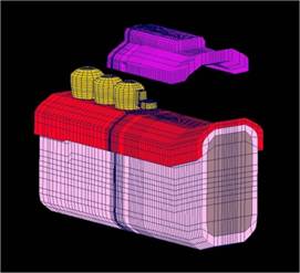
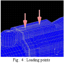
Analysis range is between the lower canine to the retromolar pad under the assumption that the teeth #6 and #7 were missing, and “ GIGAUSS C600 EC Keeper Tray ( GC ) ” was used as an extracoronal attachment ( Fig. 1 ). Contact conditions were introduced between the attachment and a denture, and the mucosa and the denture. The extracoronal attachment part was built into the model from the CAD data.
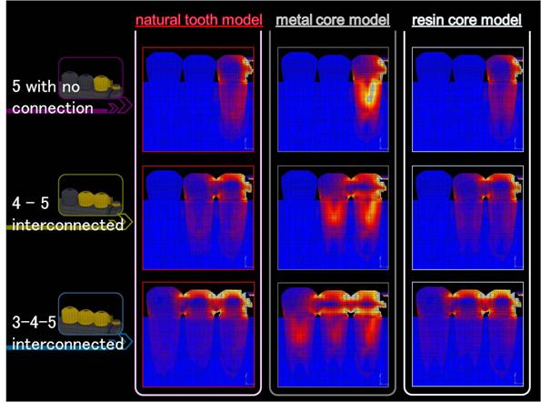
Fig. 2 shows the mesial - distal cross sectional view and components of the model. Three models including natural tooth model ( natural teeth were used as abutments ), metal core model ( metal cores were attached on abutments ), and resin core model ( resin cores were attached on abutments ) were fabricated as analytical model. The table shows the components of the model. The post length is about 2 / 3 of the root length and about 1 / 3 of the root diameter.
![]()
Fig . 1 : Overview of the model
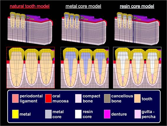 |
Fig . 2 : Compornents of each model
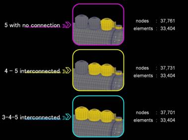
# 5 single abutment tooth, # 4 - # 5 interconnected abutment teeth, and # 3 - # 4 - # 5 interconnected abutment teeth with simplified designs were fabricated on the three models, and the effect of abutment teeth connection was compared ( Fig. 3 ).
 |
||||||
|
||||||
|
|
||||||
2 . Analysis condition
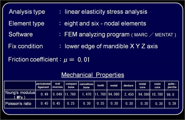
Table. 1 shows the analysis condition and mechanical properties. Analysis type was the elastic stress analysis, and general finite element programMARC / MENTAT ( MSC Software, Japan ) was used for the model fabrication and the analysis. The restraint condition was a full restraint at the lower edge of the mandible XYZ axis.
 |
|
Table . 1 : Analysis condition and mechanical properties
3 . Loading condition
Loading points are central fossae of the first and second molars. Vertical loads of 10 N at each point, and a total of 20 N were applied on the occlusal surface ( Fig. 4 ).
 |
Results
1 . Stress distribution
Fig. 5 is a mesial - distal crow sectional view through the tooth axis illustrating the stress distribution of each model.
In a # 5 single abutment tooth group, stress was observed all around the root surface, especially around the post.
In # 4 - # 5 interconnected abutment teeth group, stress concentration at the post and surrounding area was mitigated as compared to a single abutment tooth group.
In # 3 - # 4 - # 5 interconnected abutment teeth group, each model demonstrated stress mitigation, but metal core model showed higher stress concentration around the abutment teeth compared to the natural tooth model.
 |
![]()
Focusing on # 5 which is the most distal abutment tooth, there is a tendency that stress at the root was mitigated with the increase of the number of abutment teeth. In # 3 - # 4 - # 5 interconnected abutment teeth group, st interconnected ress concentration around the abutment teeth was thelargest in the metal core model.
Fig. 6 - a shows stress values at nodal points where the highest stress concentration was observed. Metal core model generally showed significantly high stress values compared to other two models although there was some effect in connecting abutment teeth.
Fig. 6 - b shows stress values of the compact bone around the cervical area of the most distal abutment tooth. Measuring point is as shown in thefigure. No significant difference was observed among each model although stress values decreased with an increase in the number of abutment teeth.
2 . Displacement
1) Displacement of each abutment tooth
Fig. 7 shows the mesial - distal displacement of each abutment tooth. A crown apex of each tooth was selected as a measuring point.
As for the tooth # 3, no significant displacement was observed among the three models in a single # 5 abutment tooth and # 4 - # 5 interconnected abutment teeth groups, but distal displacements were observed in the # 3 - # 4 - # 5 interconnected group ( Fig. 7 - a ).
As for the tooth # 4, displacements were the largest in the # 4 - # 5 interconnected group, and were decreased in the # 3 - # 4 - # 5 interconnectedgroup ( Fig. 7 - b ).
The tooth # 5, which is the most distal abutment tooth, showed the largest
displacement in a # 5 single abutment tooth group, and showed thesmallest displacement in the # 3 - # 4 - # 5 interconnected group ( Fig. 7 - c ).
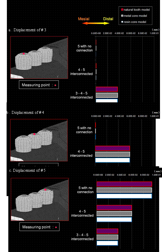
Fig . 7 : Mesial – distal displacement of each abutment toot
2)
Displacement
of dentures
Fig. 8 shows the subsidence of the denture. The measuring point is as shown in the figure. The subsidence was the largest in a # 5 singleabutment tooth group, and was the smallest in # 3 - # 4 - # 5 interconnected abutment teeth group.
|
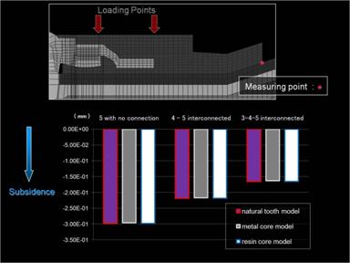
Fig..8 : Vertical displacement of denture
Discussions
Little difference was observed in displacement and stress values of the compact bone among the three models. The vertical displacement of the most distal abutment tooth and the vertical settling of a denture decreased with the increase in the number of interconnected abutment teeth. However, metal core models did not show significant stress mitigation with the increase in the number of interconnected abutment teeth.
Conclusions
The connection of remaining abutment teeth is essential in unilateral free-end edentulous cases. It is clinically important to examine the condition of abutment teeth prior to decisions regarding the selection of number of connected abutment teeth. In the present study, no significant difference was observed in stress values of the compact bone around the cervical region, mesial- distal displacement of each abutment tooth, and the denture settling, but stress in the root was significantly high in metal core models. This result suggests that more teeth should be connected to achieve the most correct tooth connection effect in metal core models compared with natural tooth models.
References
1. Tanaka, Y.: Dental Magnetic Attachment - new prothodontic treatment used the magnet -, Ishiyaku Publishers, Inc. (Tokyo), 1992.
2. Tanaka, Y.: Magfit System - the main point of clinical utilization -,DentalDiamond Co. (Tokyo), 1993.
3. Tanaka, Y.: Dental Magnetic Attachment, Q&A, Ishiyaku Publishers, Inc. (Tokyo), 1995.