The Effect of the Angle of Axial Surface of Root Cap upon the Abutment Tooth During Simulated Incisal Clenching –The Boundary Condition of Considering Human Mandible Movement–
D.
Yamanaka1, T. Ohyama 1,2,
1Department
of Partial Denture Prosthodontics,
2Division
of Clinical Research,
Introduction
When the root cap is applied to the abutment tooth for an overdenture, the shape of the root cap may affect the stress distribution of an abutment tooth and the circumferential tissue. In the case of anterior residual teeth, the clenching on residual teeth has an influence for the abutment teeth and the residual ridge. In this study, Incisal clenching was simulated newly with the joint element which permitted rotations and translations, and it was applied on the mandibular condyle and bite points on complete overdentures. It was analyzed the effects of the axial surface angle of root cap for the abutment teeth and the circumferential tissue using three-dimensional finite element analysis.
Materials and Methods
A complete overdenture model with root caps delivered on mandibular bilateral canine was evaluated. The outline of
the abutment teeth and mandible were modeled on the basis of data from a
multi-detector CT (Asteion Super4 Edition,
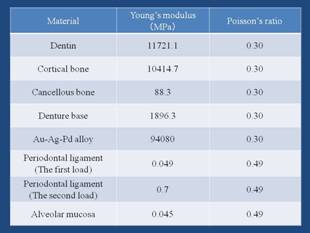
Table 1: Material Properties
The height of the root cap was 2.5mm from the lingual alveolar crest, and the top surface was set parallel to the occlusal plane. Three inclination angle (0,15, and 30 degrees) on the axial surface of the root cap were designed (Fig.1).
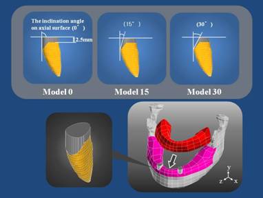
Fig.1: The Design of Root Cap
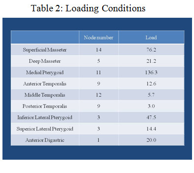
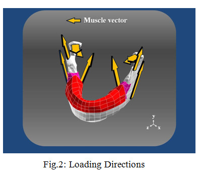
The loading condition set up the vector and force of muscular contraction of the incisal clenching.
Table 2 shows the loading conditions. Figure 2 shows the loading directions with arrows.
The joint element was
applied on the upperpart of the mandibular
condyle and sixteen occlusal
stops on complete overdenture. In modeling of joints between two
parts, the joint element permitted simple kinematic constraints such as
identical displacements between the two parts at the junction or more
complicated kinematic constrains that allow for transmission of motion between
two flexible bodies (Fig.3).
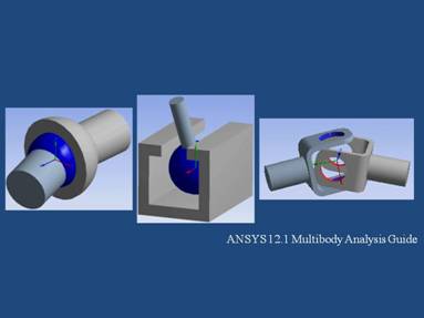
Fig.3: The Types of Joint Element
The paths of each mandibular condyle were constrained. Guidance by the articular eminence was simulated with a planar constraint. It permitted rotations in all dimensions and translations in the specified plane, and was analogous to a sphere moving between two parallel, frictionless flat plates.
The constraining plane was angled 30°forward and downward relative to the horizontal dental occlusal plane, and it was canted 5°medially. The inclination angle of cusp of artificial teeth was set 25°and balanced occlusion was simulated by sixteen sixteen occlusal stops on complete overdenture (Fig.4).
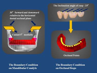
Fig.4: Boundary Conditions
Stress levels were
calculated under the minimum principal stress on the surface of cortical bone. And the force added to the abutment
teeth were calculated on the nodes of the surface of the abutment teeth.
Results
Figure 5 shows the stress distribution of the abutment teeth. Figure 6 shows the stress distribution graph of the top surface of cortical bone. The stress concentration was detected on the labial and lingual side of the abutment teeth. The stress concentration was detected on the anterior surface in comparison with the posterior surface of cortical bone, and in the model of which inclination angle of the axial surface set 30 degrees the minimum principal stress showed the smallest value.
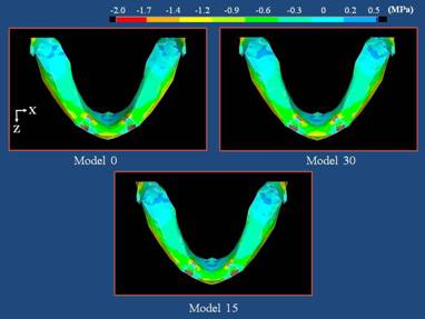
Fig.5: The Stress Distribution (Minimum Principal Stress)
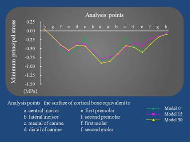
Fig.6: The Stress Distribution Graph of The Top Surface of Cortical Bone
Fig.7 shows the force added to abutment teeth during simulated incisal clenching. The force added to the abutment teeth decreased when inclination angle of the axial surface was inclined.
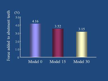
Fig.7: The Force added to Abutment Teeth
Discussions
The biomechanics of human jaw during incisal clenching was largely unknown, but in this study, the boundary condition that was similar to the human mandible enabled the biomechanical analysis during incisal clenching by applying the joint element. It is thought that the movement of the abutment teeth for the complete overdenture increased, when inclination angle of the axial surface was inclined. As a result, the force added to the abutment teeth decreased and the stress concentration was detected on the anterior surface of cortical bone.
Conclusions
By the inclination angle of the axial surface was inclined, the stress concentration was detected on the anterior surface in comparison with the posterior surface of cortical bone. However, the force added to the abutment teeth decreased.
References
1. Korioth TW, Hannam AG, Deformation of the Human Mandible During Simulated Tooth Clenching, J Dent Res 73:56-66, 1994.
2. G.E.J. Langenbach, A.G. Hannam, The Role of Passive Muscle Tensions in A Three-dimensional Dynamic Model of the Human Jaw, Arch Oral Biol 44:557-573, 1999