Longitudinal Study of Magnetic Attachments
– Investigation of Probing Depth on Abutment
Teeth Part.2 –
M. Miwata , R. Ito , K. Hoshiai , Y. Tanaka , T. Ishigami*, K. Ishibasi**, E. Bando***, H. Sasaki****, H. Mizutani***** and T. Hosoi******
Department of Removable Prosthodontics , School of Dentistry, Aichi-Gakuin University
*Department of Partial Denture Prosthodontics , Nihon University School of Dentistry
**Department of Fixed Prosthodontics , School of Dentistry , Iwate Medical University
***Department of Fixed Prosthodontics , Institute of Health Biosciences Graduate School The University of Tokushima ****Sasaki Dental Clinic
*****Graduate School of Medical and Dental Sciences, Tokyo Medical and Dental University
******Department of Removable Prosthodontics, Tsurumi University School of Dental Medicine
Introduction
Magnetic attachments have been applied clinically in various cases. It’s useful to carry out postoperative investigations and to confirm results, as it shows the criterion of the clinical application1). Thus, magnetic attachments can be used safely. The present paper reports in an investigation of prospective magnetic attachments that has been carried out since 2003 at the Japanese Society of Magnetic Applications in Dentistry. 2)(JSMAD)
Methods
The transition of probing depth (PD) of abutment teeth was measured, and the changes of the conditions of periodontal tissue were evaluated. Immediately after cementation, oral conditions were recorded by use of an original questionnaire, and PD was measured using a 6- points method. After 5 years, patients were recalled and PD was measured once again. This time, 7departments of the JSMAD, represented by the authors of this paper, participated in this study. At the beginning of the study, there were 70 patients participants.
However, 28 patients were censored, leaving 42 patients to participate in the study.
Results
Table. 1 details of the cases which were able to participate in the study.
The dentures included 24 maxillary plates and 21 mandibular plates .
The abutment teeth included 12 incisor teeth, 29 cuspid teeth, 25 premolar teeth and 9 molar teeth. The denture plate materials of the dentures were 23 resin plates and 22 metal plates.
The results were statistically analyzed using Willcoxon signed rank test and Mann-Whitney’s U test. The significance level was set at 0.05.
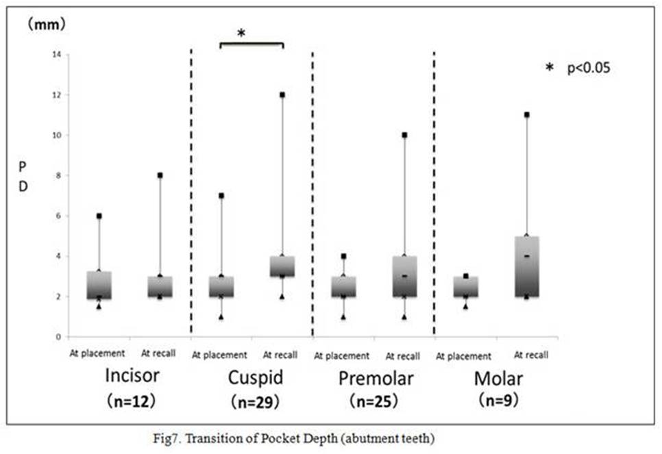
Fig 1 shows the transition with Median value and quartile deviation of PD of surviving teeth for 5 years.
A significant difference was found between initial PD and PD after 5 years of all methods (max point of olar unit, six point of olar unit, max point of all abutment teeth, six point of all abutment teeth).
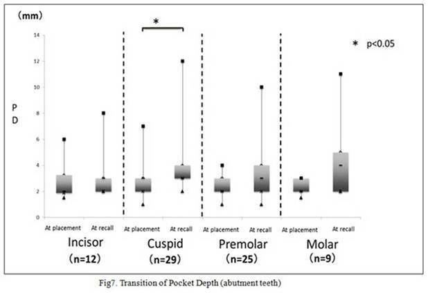
Fig 2 shows the comparison of PD between residual teeth and extracted teeth at placement.
Significant difference was found between residual teeth and extracted teeth at placement.
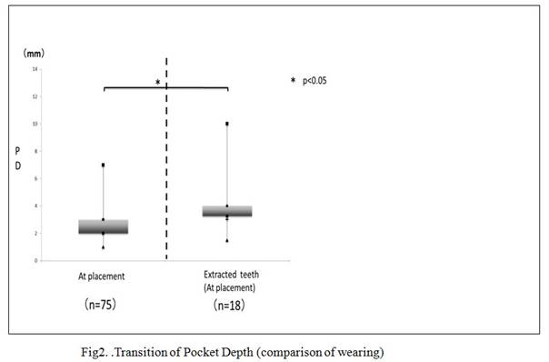
Fig 3 shows the transition of PD of maxillary and mandibular plates. Significant differences were found in the maxillary plates.
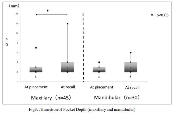
Fig 4 shows the transition of PD by gender. Significant differences were found between placement and recall for females. Significant differences were found between males and females at placement. The PD of females significantly deteriorated.
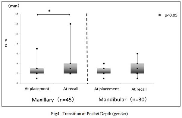
Fig 5 shows the transitions of PD of materials of plates. No significant differences were found .
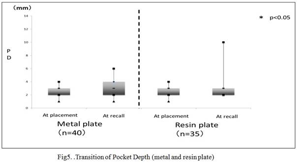
Fig 6 shows the transitions of PD of materials of plates and gender. Significant differences were found in Female/Resin plates. Significant differences were found between Female/Metal plates and Female/Resin plates at recall. Significant differences were found between Male/Metal plates and Female/Metal plates at placement.
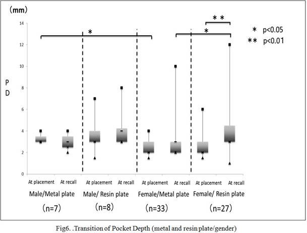
Fig 7 shows the transitions of PD by abutment teeth. Significant differences were found for cuspids.
Discussion
The null hypothesis that PD (Probing depth) of the abutment teeth would not deteriorate was rejected, as a significant difference was found between initial PD and the PD after 5 years. We reported in a retrospective cohort study that the survival rate of resin plates was worse than that of metal plates2). In the present study, the resin plate analysis did not produce the same results, and therefore, cannot be said to correspond to the cohort. However, we can say that when considering the response looking at the data for the female,s cohort, the major connector of metal plates is solid; thus, flexure of metal plates is less than that of resin plates. Therefore the abutment teeth of the metal plates were not moved, meaning that the PD of metal plates was less than that of resin plates. With respects to the significant differences found in maxillary plates and for women, further study is required.
Conclusion
The results of prospective observations of magnetic attachments from the point of the transition of PD for 5 years are given below:
・ PD of the abutment teeth were increased over a 5-year period.
・ Significant differences were found between initial PD and PD 5 years later for maxillary plates, females, Female/Resin plates, and cuspids.
・ Significant differences were found between residual teeth and extracted teeth , Male/Metal plate and Female/Metal plate of initial PD.
・ Significant differences were found between Female/Metal plate and Female/Resin plate for the PD after 5 years.
References
Literature references should be listed at the end of the paper in the same order that they appear in the text, and in accordance with the following examples.
1. K. Hoshiai, Y. Tanaka, M. Kawakita, W. Fujinami, K. Wakayama, Y. Imaizumi, T. Matumoto and M. Sakane: Longitudinal Study on Metal Plate Denture with Magnetic Attachments-Part 4, J J Mag Dent, 13(2), 26-29, 2004.
2. R. Ito, K. Hoshiai, Y. Tanaka, T. Ishigami, K. Ishibashi, E. Bando and H, Sasaki: Longitudinal Study of Magnetic Attachments -Investigation of Probing Depth on Abutment teeth-, J J Mag Dent, 19(2), 35-39, 2010.